


Fusobacterium necrophorum
-
General information
Taxonomy
Family: Fusobacteriaceae
2 subspecies:
F. necrophorum subsp fundiliforme (biovar B) dominant isolate in humans
F. necrophorum subsp necrophorum is a commonly pathogen in animals
Natural habitats
Mainly isolated from the oral cavity
Clinical significance
This bacterium is extremely virulent and caused very severe infections, mostly in children or young adults, coming through a pharynges tonsillitis.
It was formerly the most anaerobic bacteria found in tonsillar abscesses of young adults.
They also occur in abscesses in the lungs, pleura and liver.
F. necrophorum is now less found than in the era before the use of antibiotics, so this bacterium is very treacherous, because doctors are not familiar with F. necrophorum.
A Gram-stained smear with highly pleomorphic Gram-negative rods in a positive blood culture bottle from a septic patient following a sore throat may indicate invasive Fusobacterium necrophorum infection.
Lemierre syndrome
Develops most often after a strep sore throat has created a peritonsillar abscess, anaerobic bacteria like Fusobacterium necrophorum can flourish.
The bacteria penetrate from the abscess into the neighboring jugular vein in the neck and there they cause an infected clot (thrombosis) to form, from which bacteria are seeded throughout the body by the bloodstream (bacteremia).
Pieces of the infected clot break off and travel through the lungs as emboli blocking branches of the pulmonary artery bringing the heart’s blood to the lungs.
This causes shortness of breath, chest pain and severe pneumonia.
-
Diseases
-
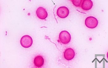
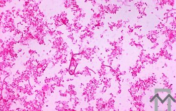
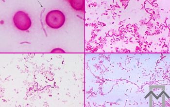
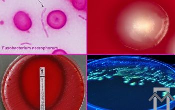
Gram stain
Pleomorphic long Gram negative rods,
with round ends and often has bizarre forms,
with sometimes round bodies.
-
Culture characteristics
-
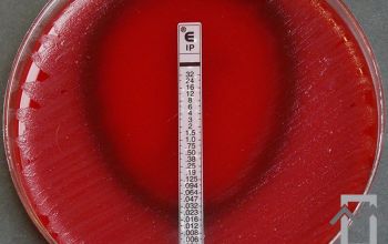
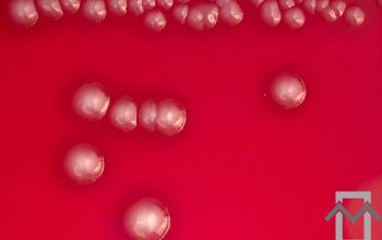
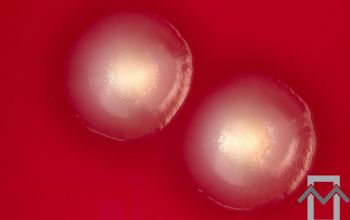
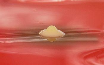
Obligate anaerobic
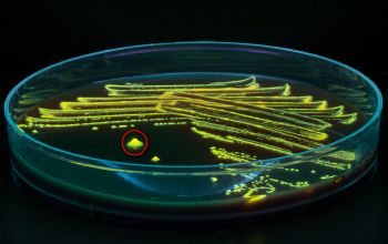
BBAØ: colonies are greyish to yellow dull, umbonate, often β-hemolytic and range in size from 0.5 to 2 mm, and they produce greening of the agar upon exposure to air.
Lipase positive strains are often β-hemolytic
BBEØ: no growth
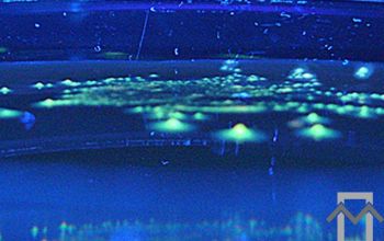
Fluorescentie/Woodslamp chartreuse / yellow-green
-
-
Characteristics
-
References
James Versalovic et al.(2011) Manual of Clinical Microbiology 10th Edition
Karen C. Carrol et al (2019) Manual of Clinical Microbiology, 12th Edition
Wadsworth-KTL, Anaerobic Bacteriology Manual, sixth edition, Hannele R. Jousimies-Somer, etc
Microbiology on the go. An initiative by
Dept. Medical Microbiology and Infectious diseases