
-350x220.jpg)
Rat Bite Fever and Streptobacillus moniliformis
Sean P. Elliot
Clinical Microbiology Reviews
Jan. 2007.p.13-22
http://cmr.asm.org/content/20/1/13.figures-only
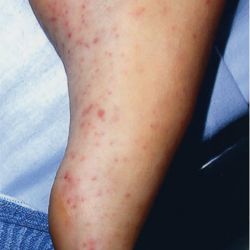
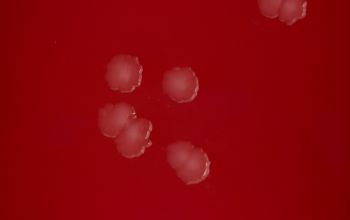
petechial and purpuric lesions on the foot of a rat bite fever patient
Sean P. Elliot
Clinical Microbiology Reviews
Jan. 2007.p.13-22
http://cmr.asm.org/content/20/1/13.figures-only
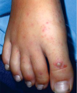
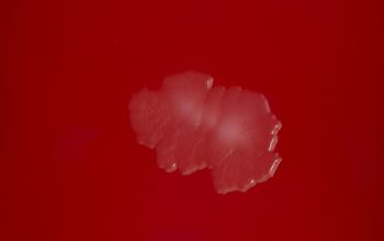
hemorrhagic vesicles on the first and third toes of a patient with advanced rat bite fever
Sean P. Elliot
Clinical Microbiology Reviews
Jan. 2007.p.13-22
http://cmr.asm.org/content/20/1/13.figures-only
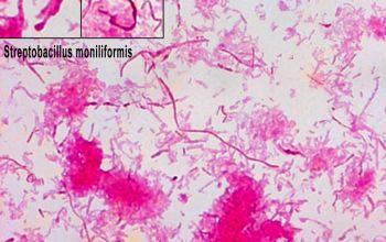
Streptobacillus moniliformis
-
General information
General Information
This organism is known to spontaneously develop L forms (bacteria without cell walls) which may allow its persistence in some sites
Taxonomy
Family: Leptotrichiaceae
Natural habitats
In the upper respiratory tract of rats (mice, gerbils, squirrels, ferrets, weasels and other rodents)
Clinical significance
They are pathogenic for humans and are transmitted by 2 routes
1. rat bite or contact with rat feces or saliva
►Rat-bite fever
2. ingestion of contaminated food
► Haverhill fever
The patient develop acute onset of chills, fever, headache, vomiting and often severe joint pains.
Complication can occur, including endocarditis, brain abscess, prostatitis and pancreatitis
-
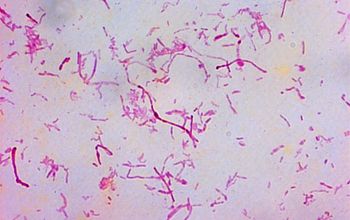
Gram stain
Gram negative rods
occuring singly or in long wavy chains or filaments (up to 150 µm).
Some may be highly pleomorphic.
Singly rods may show central swelling.
Chains or filaments may have a series of swellings (1-3 µm wide) resulting in a "string of beads" appearance.
0.1-0.7 x 1.0-5.0 µm with rounded or pointed ends.
L-forms
are coccobacillary or bipolar-staining coccoid forms, usually a special stain is required because of their lack of cell wall
(Giemsa, acridine orange)
-
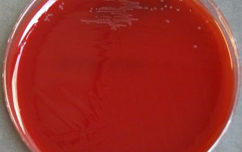
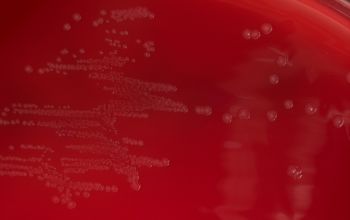
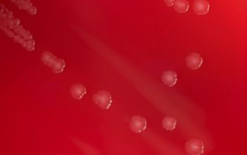
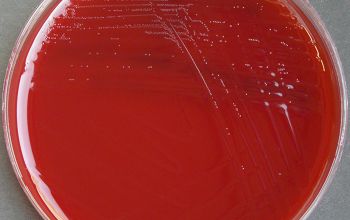
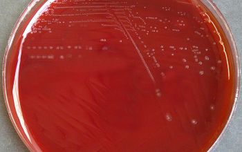
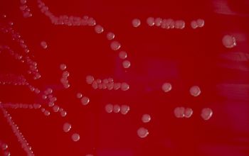
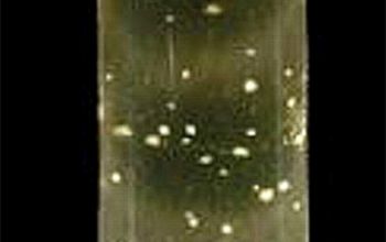
Culture characteristics
-
Facultative anaerobic (capnophilic)
They require serum, ascitic fluid and blood for growth and incubated with 5-10% CO2
BA: they grow slowly (2-3 days) and that may take as long as 7 days.
The colonies are small, circular, convex, grayish, smooth, and glistening.
L-forms: may develop in the same culture (after 5 days) are considered not pathogenic
These colonies are embedded in the agar and may also have a “fried egg” appearance, with a dark center and a flattened lacy edge.
They have undergone spontaneous transformation to the L-form.
McC: no growth
Thioglycollate broth + 10-30% ascitic fluid:
Growing colonies as "bread crumbs" on the bottom of the tube.
Blood Culture Media with SPS inhibits the growth
-
-
Characteristics
-
References
James Versalovic et al.(2011) Manual of Clinical Microbiology 10th Edition
Karen C. Carrol et al (2019) Manual of Clinical Microbiology, 12th Edition
Rat Bite Fever and Streptobacillus moniliformis Sean P. Elliot Clinical Microbiology Reviews Jan. 2007.p.13-22
Microbiology on the go. An initiative by
Dept. Medical Microbiology and Infectious diseases