
Erysipeloid is an occupational disease, occurring most frequently among veterinarians, butchers, and particularly fish handlers
The lesions usually violaceous and painful, indurated with edema inflammation but without suppuration, and clearly delineated at the border.
Dissemination and endocarditis can occur, especially in immunocompromised patients; their prognosis is generally poor.
Healing of erysipeloid usually takes 2-4 weeks, sometimes months, and relapses are common.
Author Dr. Jan R. Mekkes. Dermatoloog, AMC Amsterdam
http://www.huidziekten.nl/zakboek/dermatosen/etxt/Erysipeloid.htm
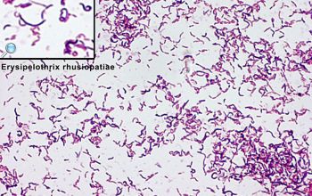
Erysipelothrix rhusiopathiae
-
General information
Taxonomy
Family: Erysipelotrichaceae
Non pathogenic: E. tonsillarum and E. inopinata
Natural habitats
Is distributed worldwide in nature, and is carried by a variety of animals, mammals, birds and fish but is most frequently associated with pigs.
Contamination of water and soil from the feces and urine of sick and asymptomatic animals often occurs.
Clinical significance
They causes mostly erysipeloid, a localized cellulitis within 2-7 days around the inoculation side.
The disease is contacted through the skin abrasion, injury, or a bite on the hands or arms of individuals handling animals or animal products.
They include peritonitis, endophtalmitis, osteomyelitis, intracranial and spinal abscesses, prosthetic joint arthritis, pneumonia, and meningitis.
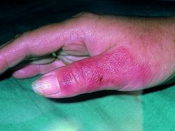
Erysipeloid
is an occupationaldisease, occurring most frequently among veterinarians, butchers, and particularly fish handlers.
The lesions usually violaceous and painful, indurated with edema inflammation but without suppuration, and clearly delineated at the border.
Dissemination and endocarditis can occur, especially in immunocompromised patients; their prognosis is generally poor.
Healing of erysipeloid usually takes 2-4 weeks, sometimes months, and relapses are common.
Lesions should be taken for Gram-staining and culture.
SWABS from the surface of the skin are not useful.
-
Diseases
-
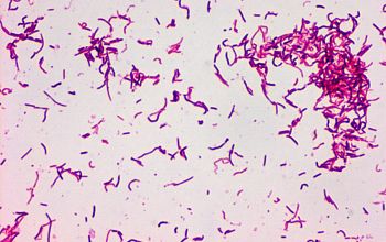
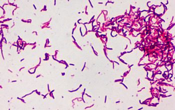
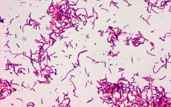
Gram stain
Short Gram positive rods,
0.2-0.5 x 0.8-2.5 µm,
occur singly, in short chains,
or in long filaments, 60 µm or more in length.
Some cells stain unevenly
There are two types
1 "smooth": small, slender rods or coccoid rods, sometimes they form short chains
2 "rough" : thin, filamentous Gram-positive rods, often more than 60 µm or more in length.
Some cells stain unevenly, which have a tendency to decolorization, whereby they are gram negative.
Because of this variation in staining and colony morphology, wrongly gave the impression that it is a matter of a polymicrobiele infection.
Isenberg
Tissue: long, slender Gram postive rods
Blood: small "corynevorm" rods
-
Culture characteristics
-
Facultative anaerobic
CO2 stimulates growth
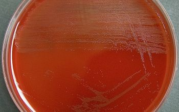
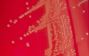
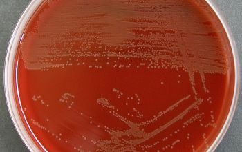
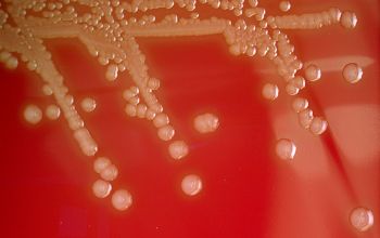
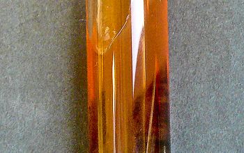
BA: after 24 h appear very small (pinpoint) colonies (<0.1-0.5)
After 48 h 2 distinct colony types
1 "smooth" sphere, round, translucent, very small, and with an entire edge, 0.3-1.5mm
2 "rough" slightly larger and flatter, with a matt surface and with an irregular edge.
A zone of greenish discoloration frequently develops underneath the colonies on blood agar plates after 48 h of incubation, making them look like colonies of viridans streptococci.
McConkey: no growth
BBAØ: growth
-
-
Characteristics
- Gram-positive
- bacilli
- growth both-aerobic-and-anaerobic
- no growth on MacConkey agar
- alpha haemolysis
- catalase-negative
- oxidase-negative
- indole-negative
- urease-negative
- TSI production of H2S
- nonmotile
- vancomycin-resistant
- colistine-resistant
- Gram positive rod- tissue-long and slender
- Gram positive rod-blood-small corynevorm
-
References
James Versalovic et al.(2011) Manual of Clinical Microbiology 10th Edition
Karen C. Carrol et al (2019) Manual of Clinical Microbiology, 12th Edition
http://www.huidziekten.nl/zakboek/dermatosen/etxt/Erysipeloid.htm
Microbiology on the go. An initiative by
Dept. Medical Microbiology and Infectious diseases